Home > Popular Themes > Human Body
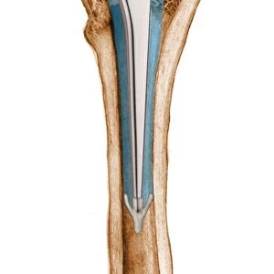
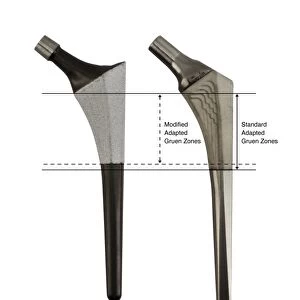
Prosthetic hip joint and Gruen zones C016 / 6779

![]()
Wall Art and Photo Gifts from Science Photo Library
Prosthetic hip joint and Gruen zones C016 / 6779
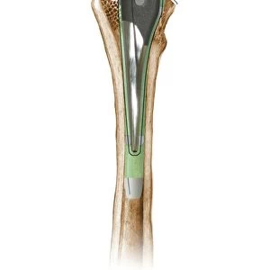
Prosthetic hip joint and Gruen zones. Cutaway diagram of a femur (thigh bone) showing a femoral component of a hip prosthesis and labels indicating Gruen zones. The femoral component is implanted in the femur after the head of the femur has been surgically removed. The other components of the hip joint are a rounded ball to fit into the socket implanted in the patients pelvis (not shown). Gruen zones are used to assess bone mineral density (BMD) after total hip replacement surgery. This is an Omnifit prosthesis fitted without bone cement. For a set of Gruen zone diagrams, see C016/6776 to C016/6781
Science Photo Library features Science and Medical images including photos and illustrations
Media ID 9243249
© D & L GRAPHICS / SCIENCE PHOTO LIBRARY
Arthritic Arthritis Arthrology Arthroplasty Artificial Bioceramic Bone Mineral Density Cutaway Device Diagram Femoral Femoral Shaft Femur Hip Implant Hip Replacement Hip Revision Implanted Internal Joint Label Labeled Labelled Labels Metal Offset Orthopaedic Orthopaedics Orthopedic Orthopedics Osteoarthritis Osteological Osteology Perimeter Profile Prostheses Prosthesis Prosthetic Prosthetics Repair Repaired Replacement Shaft Surgery Surgical Total Hip Replacement Treated Treatment Cutouts Section Sectioned
EDITORS COMMENTS
This print showcases a detailed illustration of a prosthetic hip joint and Gruen zones. The cutaway diagram reveals the inner workings of this remarkable medical device, specifically highlighting the femoral component implanted in the thigh bone after surgical removal of its head. While other components, such as a rounded ball fitting into the pelvic socket, are not shown in this image, their presence is implied. Gruen zones play a crucial role in assessing bone mineral density (BMD) following total hip replacement surgery. These labeled zones provide valuable information about the effectiveness and longevity of the prosthesis within the patient's body. The absence of bone cement further emphasizes the advanced technology employed by this Omnifit prosthesis. The artwork beautifully captures both the intricate design and functionality of this artificial joint. Its inclusion serves as an educational tool for orthopedic surgeons and medical professionals who specialize in hip replacements or revisions. With its clean white background contrasting against metal elements, this print exudes professionalism while highlighting key aspects related to treatment options for individuals suffering from arthritis or other arthritic conditions affecting their hips. By focusing on internal structures and labeling essential parts, it offers viewers an insightful glimpse into how modern medicine utilizes technological advancements to repair and restore mobility within our bodies.
MADE IN THE USA
Safe Shipping with 30 Day Money Back Guarantee
FREE PERSONALISATION*
We are proud to offer a range of customisation features including Personalised Captions, Color Filters and Picture Zoom Tools
SECURE PAYMENTS
We happily accept a wide range of payment options so you can pay for the things you need in the way that is most convenient for you
* Options may vary by product and licensing agreement. Zoomed Pictures can be adjusted in the Cart.