Prosthetic hip joints and Gruen zones C016 / 6781

![]()
Wall Art and Photo Gifts from Science Photo Library
Prosthetic hip joints and Gruen zones C016 / 6781
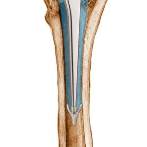
Prosthetic hip joints and Gruen zones. Diagram of the femoral components of two types of hip prosthesis with labels indicating standard and modified Gruen zones. The components are implanted in the femur (thigh bone) after the head of the femur has been surgically removed. The other components (not shown) are a rounded ball to fit into the socket implanted in the patients pelvis. Gruen zones are used to assess bone mineral density (BMD) after total hip replacement surgery. These are both cementless prostheses (Symax at left). For a set of Gruen zone diagrams, see C016/6776 to C016/6781
Science Photo Library features Science and Medical images including photos and illustrations
Media ID 9245877
© D & L GRAPHICS / SCIENCE PHOTO LIBRARY
Adapted Arthritic Arthritis Arthrology Arthroplasty Artificial Bioceramic Bone Mineral Density Comparing Comparison Device Diagram Femoral Femoral Shaft Femur Hip Implant Hip Replacement Hip Revision Joint Label Labeled Labelled Labels Metal Modified Offset Orthopaedic Orthopaedics Orthopedic Orthopedics Osteoarthritis Osteological Osteology Perimeter Profile Prostheses Prosthesis Prosthetic Prosthetics Repair Repaired Replacement Shaft Standard Surgery Surgical Total Hip Replacement Treated Treatment Cutouts Section Sectioned
EDITORS COMMENTS
This print showcases prosthetic hip joints and Gruen zones, providing a detailed diagram of the femoral components of two types of hip prosthesis. The image highlights the standard and modified Gruen zones with labels, which are essential in assessing bone mineral density (BMD) after total hip replacement surgery. These cementless prostheses, including Symax on the left side, are ingeniously designed to be implanted in the femur following surgical removal of the head of the femur. The photograph emphasizes both the technological advancements and intricate nature of artificial joint replacements. It serves as a visual representation of medical innovation and expertise in orthopedic surgery. With its white background and metal elements, this artwork provides an insightful perspective into how these devices aid in repairing damaged hips caused by conditions such as osteoarthritis or injury. The labeled sections offer a comprehensive comparison between different components, enabling healthcare professionals to make informed decisions when selecting appropriate implants for patients undergoing hip revision or total hip replacement procedures. This image is not only informative but also visually striking due to its cut-out style that allows viewers to examine each element closely. Overall, this print from D & L GRAPHICS / SCIENCE PHOTO LIBRARY captures both the technical aspects and aesthetic appeal associated with prosthetic hip joints while highlighting their crucial role in improving quality of life through advanced medical interventions.
MADE IN THE USA
Safe Shipping with 30 Day Money Back Guarantee
FREE PERSONALISATION*
We are proud to offer a range of customisation features including Personalised Captions, Color Filters and Picture Zoom Tools
SECURE PAYMENTS
We happily accept a wide range of payment options so you can pay for the things you need in the way that is most convenient for you
* Options may vary by product and licensing agreement. Zoomed Pictures can be adjusted in the Cart.