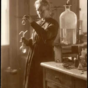
Heart anatomy, 19th Century illustration

![]()
Wall Art and Photo Gifts from Science Photo Library
Heart anatomy, 19th Century illustration
Heart anatomy, 19th Century illustration. Historical hand coloured lithographic print showing the anatomy of the human heart and the coronary arteries (thin red) that supply it. The heart is composed of four chambers. Two upper chambers, the atria (upper centre), and two lower chambers, the ventricles (lower centre). The Aorta (red tube, upper right) carries oxygenated blood from the left ventricle to the body. Image from Traite complet de l anatomie de l homme, comprenant la medecine operatoire Vol. 4 (1836), by Jean-Baptiste Marc Bourgery and illustrated by Nicolas-Henri Jacob
Science Photo Library features Science and Medical images including photos and illustrations
Media ID 6326487
© SCIENCE PHOTO LIBRARY
1836 Aorta Arteries Cardiac Circulatory Descriptive Anatomy Diagram Exterior External French Human Heart Lithograph Lithographic Print Organs Pulmonary Artery Vascular System Veins Ventricle Vessels Vol 4 Volume Four Blood Supply Cardiovascular System Circulation Plate 9 Vein
MADE IN THE USA
Safe Shipping with 30 Day Money Back Guarantee
FREE PERSONALISATION*
We are proud to offer a range of customisation features including Personalised Captions, Color Filters and Picture Zoom Tools
SECURE PAYMENTS
We happily accept a wide range of payment options so you can pay for the things you need in the way that is most convenient for you
* Options may vary by product and licensing agreement. Zoomed Pictures can be adjusted in the Cart.