Home > Animals > Mammals > Nesomyidae > Fat Mouse
Human neck anatomy, artwork C017 / 7259

![]()
Wall Art and Photo Gifts from Science Photo Library
Human neck anatomy, artwork C017 / 7259
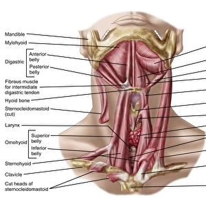
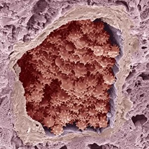
Human neck anatomy, artwork. This dissection of the neck shows the larynx (voicebox) at the upper end of the trachea (windpipe, blue). These structures have been removed and are at lower left, including the thyroid gland (yellow) that partially encloses the larynx. Tissues within the neck in the main artwork include muscles (red bands), nerves (yellow strands), veins (blue), arteries (bright red), and fat layers (yellow). Partially seen, revealed by the removal of the trachea, is the oesophagus (gullet, red with white rings of cartilage). The two main blood vessels are the internal jugular vein and the external carotid artery of the left-hand side of the neck
Science Photo Library features Science and Medical images including photos and illustrations
Media ID 9234467
© JOSE ANTONIO PE
Arteries Cartilage Dissected Dissection External Carotid Artery Gullet Internal Jugular Vein Larynx Muscles Muscular Neck Nerves Oesophagus Thyroid Gland Tissue Trachea Veins Vessel Voicebox Windpipe Cutouts
FEATURES IN THESE COLLECTIONS
> Animals
> Mammals
> Nesomyidae
> Fat Mouse
EDITORS COMMENTS
This print showcases the intricate anatomy of the human neck, as depicted in artwork C017 / 7259. The dissection reveals a detailed view of the larynx, commonly known as the voicebox, positioned at the upper end of the trachea or windpipe. In this image, both structures have been carefully removed and can be seen at the lower left corner. Adjacent to the larynx is the thyroid gland, partially enclosing it with its distinct yellow coloration. The main artwork highlights various tissues within the neck region, including muscles represented by vibrant red bands, delicate nerves depicted as yellow strands, veins portrayed in shades of blue, arteries vividly displayed in bright red hues, and layers of fat illustrated in a soft yellow tone. By removing sections of tissue and organs such as the trachea (windpipe), an additional glimpse into this complex anatomical landscape is revealed - specifically showcasing part of the oesophagus or gullet characterized by its red coloration and white rings made up of cartilage. Prominently featured are two crucial blood vessels: on one side we see an internal jugular vein while on another side there is an external carotid artery. This structure represents a male individual against a clean white background. Overall, this stunning illustration provides invaluable insight into human neck anatomy for educational purposes without any commercial use intended.
MADE IN THE USA
Safe Shipping with 30 Day Money Back Guarantee
FREE PERSONALISATION*
We are proud to offer a range of customisation features including Personalised Captions, Color Filters and Picture Zoom Tools
SECURE PAYMENTS
We happily accept a wide range of payment options so you can pay for the things you need in the way that is most convenient for you
* Options may vary by product and licensing agreement. Zoomed Pictures can be adjusted in the Cart.