Coagulation factor complex molecule C014 / 0410

![]()
Wall Art and Photo Gifts from Science Photo Library
Coagulation factor complex molecule C014 / 0410
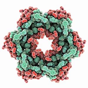
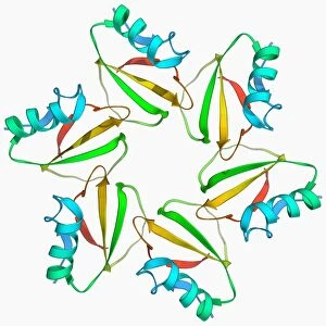
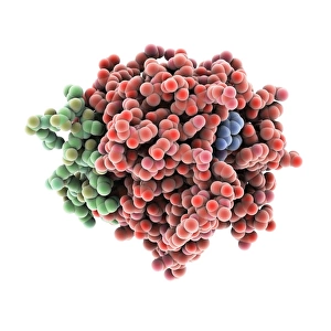
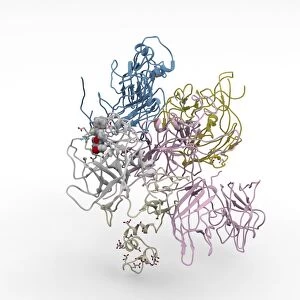
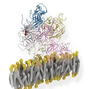
Coagulation factor complex molecule. Molecular model showing the interaction between coagulation factor VIII (FVIII, pink, blue and yellow), factor IXa (FIXa, cream and grey) and the phospholipid membrane (yellow and grey) of a blood platelet. The secondary structures (ribbons) of the molecules are shown, with the primary structure (atoms, spheres) of the active site of the factor IXa enzyme shown. These proteins are involved in the coagulation cascade, the physiological process that leads to blood clotting in the case of an injury. This complex of FVIII and FIXa activates factor X in both the extrinsic and intrinsic activation pathways
Science Photo Library features Science and Medical images including photos and illustrations
Media ID 9226837
© RAMON ANDRADE 3DCIENCIA/SCIENCE PHOTO LIBRARY
Active Site Antihaemophilic Globulin Blood Bloodstream Cell Membrane Clotting Coagulation Cofactor Complex Complexed Compound Compounds Enzyme Factor Viii Glycoprotein Interaction Membrane Platelet Secondary Structure Serine Protease Biochemical Biochemistry Fviii Molecular Model Protein Viii
EDITORS COMMENTS
This print showcases the intricate molecular structure of the coagulation factor complex molecule C014/0410. The image depicts the interaction between coagulation factor VIII (FVIII), factor IXa (FIXa), and the phospholipid membrane of a blood platelet. The vibrant colors highlight FVIII in pink, blue, and yellow, while FIXa is represented in cream and grey. The yellow and grey shades symbolize the phospholipid membrane surrounding these proteins. Ribbons illustrate the secondary structures of these molecules, while atoms and spheres depict the primary structure of the active site of the factor IXa enzyme. These proteins play a crucial role in initiating blood clotting when an injury occurs, forming part of the coagulation cascade process. This particular complex activates factor X through both extrinsic and intrinsic activation pathways. The artwork beautifully captures this essential biological mechanism that ensures our bodies can effectively respond to injuries by preventing excessive bleeding. It highlights how various compounds like glycoproteins interact with cell membranes to facilitate clot formation. With its detailed representation of biochemical interactions within our bloodstream, this image offers valuable insights into factors such as antihaemophilic globulin, hemophilia A treatment options like Factor VIII or AHF therapy, as well as other components involved in clotting processes such as serine proteases.
MADE IN THE USA
Safe Shipping with 30 Day Money Back Guarantee
FREE PERSONALISATION*
We are proud to offer a range of customisation features including Personalised Captions, Color Filters and Picture Zoom Tools
SECURE PAYMENTS
We happily accept a wide range of payment options so you can pay for the things you need in the way that is most convenient for you
* Options may vary by product and licensing agreement. Zoomed Pictures can be adjusted in the Cart.